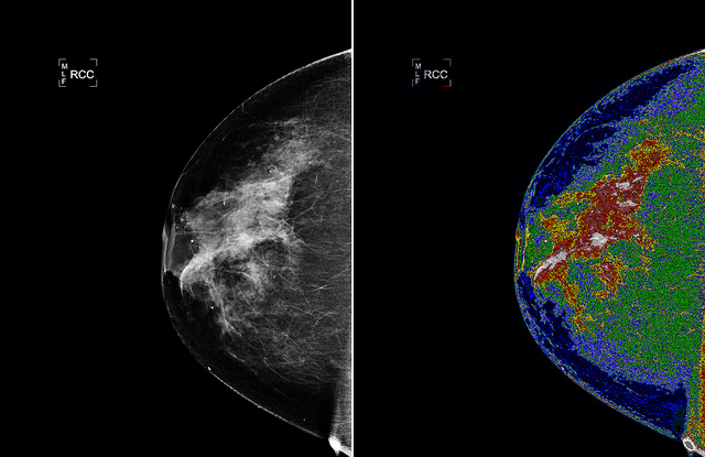
a world group comprising engineers, mathematicians and doctors has utilized a method used for detecting damage in underwater marine constructions to identify cancerous cells in breast melanoma histopathology pictures.
Their multidisciplinary leap forward, which has the talents to automate the screening of photos and enhance the detection fee, has been published in leading journal, PLOS ONE.
Breast melanoma is essentially the most customary sort of cancer for women global. current breast cancer scientific practice and treatment specially depends on the comparison of the sickness's prognosis the usage of the Bloom-Richardson grading gadget. The critical scoring is in accordance with a pathologist's visible examination of a tissue biopsy specimen below microscope, but distinct pathologists may assign distinct grades to the equal specimens.try: essential Blood examine could discover melanoma Ten Years earlier than indicators show
besides the fact that children, the advent of digital pathology and quickly digital slide scanners has opened the possibility of automating the prognosis via applying image-processing methods. whereas this absolutely represents progress, picture-processing strategies have struggled to analyse excessive-grade breast cancer cells as these cells are sometimes clustered collectively and have vague boundaries, which makes a success detection extremely challenging.
however the new method has apparently overcome that assignment, in response to Assistant Professor in Civil Engineering at Trinity college Dublin, Bidisha Ghosh. She said: "This unique analysis community could draw on a broad and deep skills base. consultants in numerical strategies and picture-processing liaised with clinical pathologists, who have been in a position to present professional insight and will tell us exactly what counsel became of cost to them. it's a superb illustration of how multidisciplinary research collaborations can address vital societal considerations."
related: New Drug evokes Hope For Alzheimer's treatment
Professor joy John Mammen, Head of branch of Transfusion medication & Immunohaematology from the Christian medical faculty, Vellore, India, spoke of: "Detection of cancerous nuclei in high-grade breast melanoma images is reasonably challenging and this work may be regarded as a primary step against automating the prognosis."
The proposed approach, in the past used for detecting broken floor areas on underwater marine structures corresponding to bridge piers, off-shore wind turbine structures and pipe-lines turned into applied to histopathology photos of breast cells. The researchers regarded the likelihood of each factor in a histopathology photo either being near a phone centre or a cell boundary. using a perception propagation algorithm, the most proper cellphone boundaries were then traced out.extra: First Ever Quadriplegic treated With Stem Cells Regains Motor control in His higher physique
This technique become developed together with mathematicians in Madras Christian school, India. Lead author, Dr Maqlin Paramanandam, said: "The advantage for this expertise is terribly interesting and we're delighted that this overseas and inter-disciplinary group has labored so neatly at tackling a true bottle-neck in automating the prognosis of breast cancer the usage of histopathology pictures."
Dr Michael O'Byrne, who additionally labored in tuition college Cork right through this project, added: "Coming from a civil engineering background where most of our image-processing equipment were designed to verify structural harm, it was fine to find some cross-over functions and find areas the place we might lend our competencies. we all discovered it specially rewarding to contribute in opposition t breast melanoma research."
(supply: Trinity faculty Dublin)
Multiply The decent: click To Share – photograph with the aid of NASA Goddard picture and Video, CC

0 komentar:
Posting Komentar